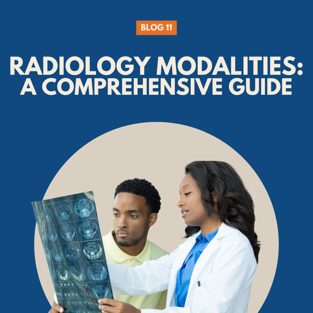
Radiology Modalities: A Comprehensive Guide
All radiology modalities play a critical role in modern healthcare, providing essential diagnostic information and guiding treatment decisions. With the advancement of technology, radiology has diversified into various modalities, each with unique capabilities and applications. We are going to explore the major radiology modalities, delving into their principles, uses, advantages, limitations, and the credentials required to operate them. Understanding these modalities can help healthcare professionals make informed decisions and improve patient outcomes.
1. X-ray Radiography
Principles
X-ray radiography is one of the oldest and most widely used radiology modalities. It involves passing X-rays, a form of electromagnetic radiation, through the body to create images of internal structures. The X-rays are absorbed at different rates by different tissues, producing a contrast image on a detector.
Applications
- Bone Fractures: X-rays are commonly used to diagnose bone fractures and dislocations.
- Chest Imaging: They are essential for detecting lung conditions such as pneumonia, tuberculosis, and lung cancer.
- Dental Imaging: X-rays help in diagnosing dental issues like cavities and impacted teeth.
Advantages
- Speed: X-ray imaging is quick, making it ideal for emergency situations.
- Cost-Effective: It is relatively inexpensive compared to other imaging modalities.
- Accessibility: Widely available in most healthcare facilities.
Limitations
- Radiation Exposure: X-rays involve exposure to ionizing radiation, which carries a risk of causing tissue damage over time.
- Limited Soft Tissue Contrast: X-rays are less effective at imaging soft tissues compared to other modalities like MRI or CT.
Credentials
- Radiologic Technologist (RT): Typically requires an associate degree in radiologic technology and certification through the American Registry of Radiologic Technologists (ARRT).
2. Computed Tomography (CT)
Principles
Computed Tomography (CT) uses X-rays to create detailed cross-sectional images of the body. The patient lies on a table that slides into a large, donut-shaped machine. The X-ray tube rotates around the patient, and detectors capture multiple views. A computer then processes these views to create detailed images.
Applications
- Trauma: CT scans are invaluable for assessing trauma patients, providing detailed images of bones, organs, and blood vessels.
- Cancer: CT is used for detecting, staging, and monitoring various cancers.
- Abdominal Issues: It helps diagnose conditions like appendicitis, kidney stones, and gastrointestinal disorders.
Advantages
- Detailed Imaging: Provides highly detailed images of bones, blood vessels, and soft tissues.
- Fast: CT scans can be completed quickly, often within minutes.
- 3D Imaging: Capable of producing three-dimensional reconstructions.
Limitations
- High Radiation Dose: CT scans expose patients to higher doses of radiation than standard X-rays.
- Cost: More expensive than traditional X-rays.
- Contrast Reactions: Some patients may have allergic reactions to contrast agents used during the scan.
Credentials
- CT Technologist: Requires certification as a radiologic technologist (RT) and additional training or certification in CT imaging through ARRT.
3. Magnetic Resonance Imaging (MRI)
Principles
Magnetic Resonance Imaging (MRI) uses powerful magnets and radio waves to produce detailed images of the body’s internal structures. Unlike X-rays and CT scans, MRI does not use ionizing radiation. It relies on the magnetic properties of hydrogen atoms in the body, which align with the magnetic field and emit signals when exposed to radiofrequency pulses.
Applications
- Neurological Imaging: MRI is the gold standard for imaging the brain and spinal cord, detecting conditions like tumors, stroke, and multiple sclerosis.
- Musculoskeletal Imaging: Used for assessing joint injuries, soft tissue tumors, and spinal disorders.
- Cardiac Imaging: Provides detailed images of the heart and blood vessels.
Advantages
- No Radiation: MRI does not use ionizing radiation, making it safer for repeated use.
- High Soft Tissue Contrast: Excellent at distinguishing between different types of soft tissues.
- Versatility: Can image nearly every part of the body in great detail.
Limitations
- Cost: MRI is more expensive than other imaging modalities.
- Time-Consuming: Scans can take 30 minutes to over an hour, requiring patients to remain still.
- Claustrophobia: The enclosed nature of the MRI scanner can be challenging for some patients.
Credentials
- MRI Technologist: Requires certification as a radiologic technologist (RT) and additional training or certification in MRI imaging through ARRT.
4. Ultrasound
Principles
Ultrasound imaging uses high-frequency sound waves to produce images of the body’s internal structures. A transducer emits sound waves that bounce off tissues and organs, and the reflected waves are captured to create real-time images.
Applications
- Obstetrics: Widely used for monitoring fetal development during pregnancy.
- Cardiology: Echocardiography assesses the heart’s structure and function.
- Abdominal Imaging: Useful for examining organs such as the liver, gallbladder, and kidneys.
Advantages
- Safety: Ultrasound does not use ionizing radiation, making it safe for pregnant women and children.
- Real-Time Imaging: Provides real-time images, useful for guiding procedures like biopsies.
- Portability: Portable machines allow for bedside imaging.
Limitations
- Limited Penetration: Ultrasound has difficulty imaging structures behind bone or gas.
- Operator Dependency: Image quality and interpretation can vary based on the operator’s skill.
- Image Quality: Lower resolution compared to CT and MRI.
Credentials
- Diagnostic Medical Sonographer: Requires an associate degree in diagnostic medical sonography and certification through the American Registry for Diagnostic Medical Sonography (ARDMS).
5. Nuclear Medicine
Principles
Nuclear medicine involves the use of small amounts of radioactive materials, or radiotracers, to diagnose and treat diseases. The radiotracer is administered to the patient, and a special camera detects the radiation emitted from the body to create images.
Applications
- Oncology: PET scans (a type of nuclear medicine) are used for detecting cancer and evaluating treatment response.
- Cardiology: Myocardial perfusion imaging assesses blood flow to the heart muscle.
- Endocrinology: Thyroid scans evaluate thyroid function and detect abnormalities.
Advantages
- Functional Imaging: Provides information about the function of organs and tissues, not just structure.
- Early Detection: Can detect diseases at an earlier stage compared to other imaging modalities.
- Targeted Therapy: Radiotracers can be used to treat certain conditions, such as thyroid cancer.
Limitations
- Radiation Exposure: Involves exposure to radioactive materials, though typically in small amounts.
- Cost and Availability: Can be expensive and not available in all healthcare facilities.
- Time: Some scans require hours or days for the radiotracer to accumulate in the target area.
Credentials
- Nuclear Medicine Technologist: Requires an associate degree in nuclear medicine technology and certification through the Nuclear Medicine Technology Certification Board (NMTCB) or ARRT.
6. Mammography
Principles
Mammography is a specialized type of X-ray imaging used to examine breast tissue. It uses low-dose X-rays to create detailed images, primarily for detecting breast cancer.
Applications
- Screening: Regular mammograms are recommended for early detection of breast cancer.
- Diagnostic: Used to investigate symptoms such as lumps or nipple discharge.
Advantages
- Early Detection: Effective for early detection of breast cancer, improving treatment outcomes.
- Detailed Imaging: Provides detailed images of breast tissue, highlighting abnormalities.
Limitations
- Radiation Exposure: Involves exposure to low doses of ionizing radiation.
- Discomfort: Compression of the breast during the procedure can cause discomfort.
- False Positives/Negatives: May lead to false-positive or false-negative results, requiring further testing.
Credentials
- Mammography Technologist: Requires certification as a radiologic technologist (RT) and additional training or certification in mammography through ARRT.
7. Fluoroscopy
Principles
Fluoroscopy is an imaging technique that provides real-time moving images of the interior of the body using X-rays. It involves continuous X-ray exposure and a fluorescent screen to create dynamic images.
Applications
- Guided Procedures: Used in guiding catheter placements, joint injections, and other interventional procedures.
- Gastrointestinal Studies: Evaluates the gastrointestinal tract, such as during a barium swallow.
- Cardiac Catheterization: Assesses the coronary arteries and heart function.
Advantages
- Real-Time Imaging: Allows for dynamic visualization of internal structures and movements.
- Guidance: Essential for guiding various medical procedures with precision.
Limitations
- Radiation Exposure: Continuous exposure to X-rays increases radiation dose to patients.
- Cost: Can be expensive, especially for complex procedures.
- Limited Soft Tissue Contrast: Less effective for detailed soft tissue imaging compared to MRI.
Credentials
- Radiologic Technologist (RT): Requires an associate degree in radiologic technology and certification through ARRT. Additional training in fluoroscopy may be required.
8. Positron Emission Tomography (PET)
Principles
Positron Emission Tomography (PET) is a nuclear medicine technique that uses radiotracers emitting positrons to create detailed images. The most commonly used radiotracer is fluorodeoxyglucose (FDG), which accumulates in areas of high metabolic activity.
Applications
- Oncology: PET is highly effective in detecting and staging cancers, monitoring treatment response, and identifying metastasis.
- Neurology: Used to study brain metabolism and diagnose conditions like Alzheimer’s disease.
- Cardiology: Assesses myocardial viability and blood flow.
Advantages
- Functional Imaging: Provides detailed information about metabolic and functional activity in tissues.
- Early Detection: Can detect abnormalities at a cellular level before structural changes occur.
- Combination Imaging: Often combined with CT (PET/CT) for precise anatomical localization.
Limitations
- Radiation Exposure: Involves exposure to radioactive materials.
- Cost: Expensive and not always widely available.
- Time: Scans can take longer due to the need for radiotracer preparation and uptake time.
Credentials
- Nuclear Medicine Technologist: Requires an associate degree in nuclear medicine technology and certification through NMTCB or ARRT. PET technologists often receive additional training specific to PET imaging.
9. Bone Densitometry (DEXA)
Principles
Dual-Energy X-ray Absorptiometry (DEXA) is a specialized form of X-ray imaging used to measure bone mineral density (BMD). It uses two X-ray beams at different energy levels to assess bone strength.
Applications
- Osteoporosis: Primarily used to diagnose and monitor osteoporosis and assess fracture risk.
- Fracture Risk Assessment: Helps in evaluating the risk of fractures in at-risk populations.
Advantages
- Accuracy: Provides accurate and precise measurements of bone density.
- Low Radiation Dose: Uses minimal radiation compared to other imaging techniques.
- Predictive Value: Effective in predicting fracture risk and guiding treatment decisions.
Limitations
- Limited Scope: Primarily focuses on bone density and does not provide detailed images of other structures.
- Access: Not always available in all healthcare settings.
- Cost: Can be relatively expensive for patients without insurance coverage.
Credentials
- Radiologic Technologist (RT): Requires certification as a radiologic technologist and additional training in bone densitometry.
Radiology modalities are indispensable tools in modern medicine, each offering unique advantages and addressing specific clinical needs. From the traditional X-ray to advanced techniques like PET and MRI, these imaging technologies provide critical information that aids in diagnosis, treatment planning, and monitoring of various medical conditions. Understanding the principles, applications, advantages, limitations, and required credentials of each modality is essential for healthcare professionals to make informed decisions and optimize patient care.
By staying informed about the latest advancements and best practices in radiology, healthcare providers can ensure that they utilize the most appropriate imaging techniques for their patients, ultimately improving outcomes and enhancing the quality of care.
At RISC Staffing, we currently are staffing the following modalities:
- Radiology
- Ultrasound
- MRI techs
We are expected to expand as time and resources allow. For more information please contact: info@riscstaffing.com
Here is a short Youtube video with more information about the different radiology modalities!


